Skip to content
Home
Skin Services
Brows
Online Brow Education
About Us
Book with us
Brow Lamination- 4 in 1 (in person)
Clinical Esthetics trainings (in person)
X
Dashboard
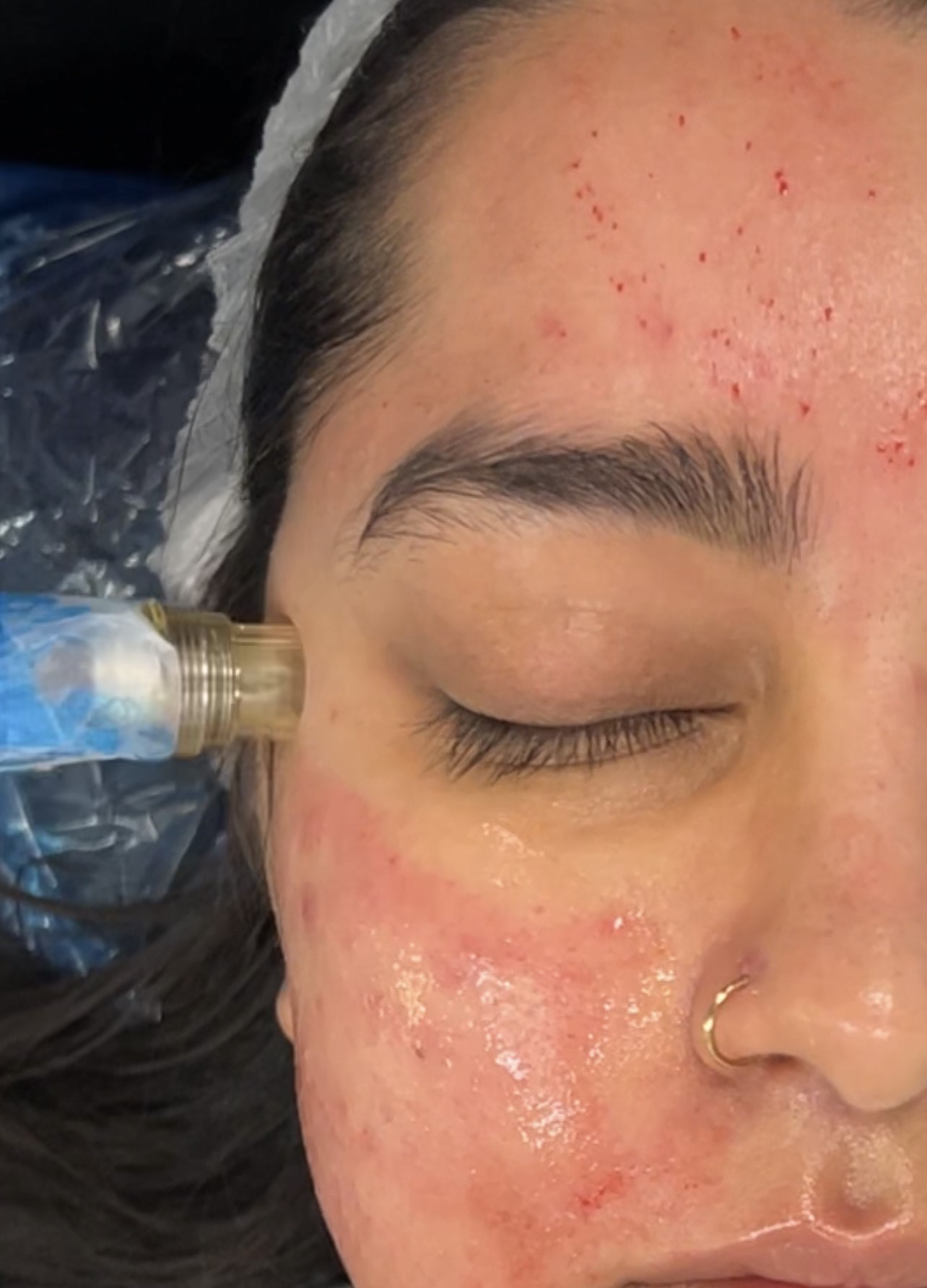
Protected: Microneedling Training (collagen induction therapy)
Uncategorized
Wishlist
Share
Share Course
Page Link
Share On Social Media
Course is locked
Protected: Microneedling Training (collagen induction therapy)
Enter your password
Show password
Submit
Login
Username or email address
*
Required
Password
*
Required
Remember me
Log in
Lost your password?